The image quality depends mostly on the ultrasound transducer, that will be the leading conclusion that communicates and gets the physical energy. Modern ultrasound transducers give users unparalled and unparalleled multi-modality ultrasound experience.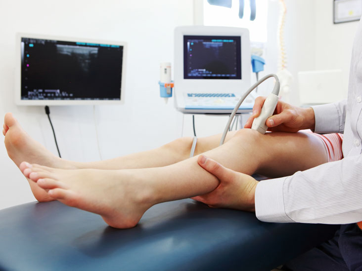
Carotid ultrasonography, applied to examine blood flow in to the carotid arteries, in addition to the intra-cerebral arteries. Echocardiography, which is an ultrasound that reveals the motion of the center, while the muscle dilates and contracts. Crisis medical experts frequently use a form of probe. Also, it’s used in the ER as a schedule way of easily assessing the explanation for a patient’s abdominal pain.
Urologists usually use ultrasound transducers to identify the degree of fluid that is in a patient’s bladder. Gynecologists may execute a pelvic sonogram utilizing an ultrasound to see an image of the pelvic floor in girls and detect any abnormalities. More in depth images of the tendons, nerves, muscles, ligaments and different smooth muscle areas that may have been affected by injury or trauma. Arterial probe is used by cardiologists to check on for probable obstructions in the arteries, or even to detect DVT. In gastroenterology, medical practioners can view the abdomen using an probe to see organs such as the aorta, pancreas, gall bladder, kidneys, liver and spleen.
There are four types of available ultrasound image. The choice that kind of image to use is dependent upon the objectives for a certain check, the phenomena being investigated and what equipment is available. The most typical and kind of ultrasound picture is a series of flat, two-dimensional combination area photos of the scanned tissue. Referred to just as second ultrasound, that function of scanning remains normal for most diagnostic and obstetric situations following a half-century of use.
Recently, 2d pictures have been predicted in to three-dimensional representations. That is accomplished by scanning structure corner parts at a variety of perspectives and reconstructing the information acquired right into a three-dimensional image. A typical use for 3d ultrasound pictures is to supply a more complete and reasonable image of a developing fetus. By upgrading 3d ultrasound images in quick sequence, sonographers also can build 4d ultrasound pictures. In the 4d ultrasound, the fourth aspect, time, adds action and generates the most realistic representation of all.
Sometimes, 3d and 4d ultrasound photographs may reveal abnormalities not easily seen applying second ultrasound. For expectant moms and family unit members, the capability to see reasonable pictures of an unborn child in the uterus can be gratifying and heartwarming although the medical community in general cautions against performing ultrasound checks only with this purpose.
Analyzing blood movement as it techniques through body ships is really a frequent component of lots of the kinds of ultrasound. While conventional second ultrasound and their three-dimensional offshoot show inner areas and structures, a different kind of ultrasound in melton is needed to examine body flow and stress inside a blood vessel. A Doppler ultrasound evaluation bounces high-frequency noise dunes down body cells in action and records changes in frequency of the sound dunes because they match back to the transducer probe. It then switches this information into a visual illustration of how quickly and in what path blood is flowing. Doppler ultrasound is definitely an indispensable diagnostic instrument in every areas of ultrasound testing and is preferable in many cases to X-ray angiography because it does not need injecting the in-patient with different dye.